
Source: image of CT scan: public domain; http://upload.wikimedia.org/wikipedia/commons/3/35/Ct-scan.jpg; CT image: public domain; http://upload.wikimedia.org/wikipedia/commons/7/7a/CT_Scan.JPG; MRI image: public domain; http://upload.wikimedia.org/wikipedia/commons/3/3b/MRI_brain.jpg?uselang=enmri; MRI:public domain; http://upload.wikimedia.org/wikipedia/commons/5/5c/Varian4T.jpg; fMRI image: public domain; http://upload.wikimedia.org/wikipedia/commons/1/15/FMRI.jpg?uselang=enmri
Hello, class. So psychologists use many different kinds of tools and techniques to map the brain out and to discover exactly how it works. So this is also used to examine specific people that have brain damage or certain disorders to uncover exactly what's going on, but it can also be used in healthy people as well to see what normal functioning of the brain is. So we're going to be examining some of those technologies and techniques in today's lesson.
So one of the first assessments of brain function and brain disorders are the physiological and behavioral changes in a person. Since the brain controls a lot of our responses, we can assume that different kinds of responses that are abnormal might be the result of different kinds of brain damage or problems with the brain function. Now these can be very obvious and profound differences.
For example, in stroke patients. They might have noticeable difficulty moving or speaking. But they can also be more subtle signs. An example of a subtle sign is a neurological soft sign, which is a term used for minor signs of nonspecific sorts of brain disorders. And these are things that might not normally be noticed by people, but which psychologists are a bit more trained to recognize and understand as signs of neurological problems.
This could be things like clumsiness or poor hand-eye coordination. Lots of things related to motor movements. And these are very helpful in diagnosing things like learning disorders, but also more profound disorders, like obsessive compulsive disorder or even schizophrenia as a sign of predicting that.
Now besides noticing the outer signs or the outward behaviors that people might show as results of neurological problems, we can also take a look at the brain structure itself. And there are specific tools that scientists and psychologists use to measure these kinds of things. And these are probably terms that you're familiar with if you've been watching any kind of crime or medical shows on TV, but we're going to talk about how they actually function.
We'll also be showing pictures that are taken with each of these instruments so you can compare and contrast them and see how they might give different images of the brain themselves. So first, let's take a look at ways that we can look at brain structure. And these are tools that either map or model the brain out, generally in 3D.
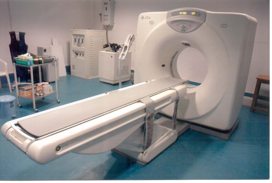
First, we have a CT scan, which is a computed tomography scan, which is essentially a specialized x-ray device. So it's taking x-rays of the brain, taking multiple images, and then creating a 3D model of the brain from those multiple images. So they can show some of the internal structures just like x-rays normally do, but it can sometimes be a bit more difficult looking inside the brain itself, looking at some of the more internal structures. For that, we need a different kind of tool like an MRI.
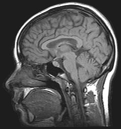
An MRI or magnetic resonance imaging device uses magnetic fields to provide more internal images of the structures of the brain in 3D as well. So both of these are 3D devices. It also doesn't use any kind of radiation, like an x-ray device, like a CT scan uses. So in that way, it's a little bit safer for the person who's using it.
There are also devices that measure the brain's function itself, which is to say it doesn't just take a map or a picture of something, but rather shows how the brain is active over time through different sorts of behaviors. So we can use it to show which areas of the brain are related to certain kinds of thoughts and actions. In this way, it's a bit more comprehensive than some of the brain structure tools.
But again, it's something that's just a little bit different. For example, if we wanted to see which areas of the brain were active when a person is dreaming we would use a functioning tool here. So one example is an EEG or an electroencephalograph, which is a device that's placed directly on a person's brain so it's not very invasive, and it can help to amplify and measure all of the brain's different electrical activity in certain areas of the brain.
So it can show the brain waves of specific areas of the brain, but they're very simple and they don't actually show much of a concrete image. Generally, just little lines on a piece of paper. To actually get a picture, we would have to use something like a PET scan or a positron emission tomography scan, which is a device that a person is put into after they've been injected with a radioactive fluid, which we call a tracer.
And then the person after this tracer has been injected into their blood does some kind of behavior or thinks about certain kinds of things. And then that radioactive tracer then goes to those specific areas that are active so it's showing where the blood flow is most prominent. And those places light up in the PET scan. And we can see that those are the areas that are being used for that certain kind of behavior.
Generally, a PET scan is used in conjunction with some kind of brain scanning, brain structure tool. Things like a CT scan. So that way you can actually get a picture along with the function itself.
But because it uses a radioactive fluid, it does require more time for that fluid to leave the system so it has to break down and go away. So you can't necessarily do this one time after the other. And it also uses a radioactive fluid so sometimes it can be a little bit dangerous with repeated usage, although the fluids they use are obviously not totally dangerous or we wouldn't use them, right?
Finally, we have a functional MRI or a functional magnetic resonance imaging device, which is similar to an MRI, but it uses those magnetic fields to measure the blood flow kind of like with a PET scan, but in a lot less invasive of a way. It doesn't use any kind of fluids. And you can see how the blood flow to certain areas over time can show how those areas are active in certain kinds of behaviors or thoughts.
In this way, there isn't any kind of known harm that these magnetic fields cause to the person's brain. So in this way, it's very useful, it's very effective, and it's a lot safer than a PET scan is as well. And it provides a lot more of a comprehensive view of the person's brain over time.